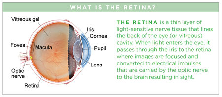
what happens to your vision when the vitreous detatches?
Posterior Vitreous Detachment
Symptoms
- Floaters (mobile blurry shadows that obscure the vision)
- Flashes (streaks of light, usually at the side of the vision)
These symptoms usually become less intense over several weeks. Most patients experience PVD after age sixty, in one case in each eye, and the condition is usually non-sight-threatening only occasionally affects vision more than permanently in the event of complication, such every bit retinal detachment or epiretinal membrane.
Symptoms in detail
Mild floaters in the vision are normal, only a sudden increase in floaters is oft the showtimesymptom of PVD.
During PVD, floaters are often accompanied by flashes, which are most noticeable in night surroundings. Most patients feel floaters and flashes during the commencement few weeks of a PVD, but in some cases the symptoms are hardly noticeable. If PVD is complicated by vitreous hemorrhage , retinal detachment , epiretinal membrane , or macular hole , the flashes and floaters may be accompanied by decreased or distorted vision. Floaters are most bothersome when virtually the center of vision and less annoying when they settle to the side of the vision. They may appear like cobwebs, dust, or a swarm of insects—or in the shape of a circumvolve or oval, called a Weiss band.
Causes
Over time, the vitreous gel that fills the heart becomes liquid and condenses (shrinks) due to age and normal wear and tear. Eventually it cannot fill the whole volume of the eye'due south vitreous cavity (which remains the aforementioned size during adulthood) and so the gel separates from the retina, located at the very back of the eye cavity.
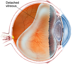
Figure i. Diagram of the vitreous cavity during posterior vitreous disengagement.
Over the next 1 to 3 months, the vitreous gel further condenses and the sides of the gel as well split up from the retina until the PVD is complete and the vitreous gel is attached to the retina only at the vitreous base (run across Effigy 1). Clear vitreous fluid fills the space between the condensed vitreous gel and the retina.
If a PVD progresses gently, gradually, and uniformly, the symptoms are typically mild. Yet, if the forces of separation are strong or concentrated in a particular part of the retina, or if there is an aberrant adhesion (sticking together) between the vitreous gel and the retina (such as lattice degeneration), the PVD tin tear the retina or a retinal blood vessel.
Flashes and floaters are typically more obvious when PVD is complicated by a retinal tear or vitreous hemorrhage. These atmospheric condition can lead to further complications, such equally retinal disengagement or epiretinal membrane, which can event in permanent vision loss. However, about 85% of patients who experience PVD never develop complications and in nearly cases, the flashes and floaters subside inside 3 months.
Risk factors
Posterior vitreous disengagement is rare in people under the age of 40, and increasingly common during avant-garde age.
Additional adventure factors for PVD include myopia (nearsighted- ness), trauma, and contempo heart surgery such equally acataract operation. Patients who experience PVD in one heart volition often experience PVD in the other center within 1 year.
Diagnostic testing
Posterior vitreous detachment is commonly diagnosed with a dilated centre examination. Notwithstanding, if the vitreous gel is very articulate, it may be hard to come across the PVD without boosted testing, such asoptical coherence tomography (OCT) orocular ultrasound (see Figure ii).
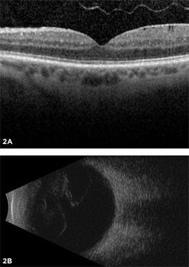
Effigy 2. Most posterior vitreous detachments can exist diagnosed with a dilated heart exam. Withal, October (A) and B-browse ultrasound (B) are diagnostic tests that can exist helpful in diagnosing PVD.
Treatment and prognosis
PVD is not-sight-threatening and the symptoms subside in the vast majority of patients. Near patients no longer notice flashes after 3 months and floaters tend to amend. No specific treatment is needed for PVD. That said, complications of PVD are rare but can be serious and require urgent treatment, such as light amplification by stimulated emission of radiation for a retinal tear or surgery for aretinal detachment. For this reason, one or more than checkups are recommended inside 3 months after the onset of PVD. In rare cases, the floaters from PVD persist, and vitrectomy surgery to remove the floaters is constructive; yous and your doc may consider this after discussing the risks and benefits of surgery.
tameztordreptles1950.blogspot.com
Source: https://www.asrs.org/patients/retinal-diseases/9/posterior-vitreous-detachment
0 Response to "what happens to your vision when the vitreous detatches?"
Post a Comment